
Fig. 1.
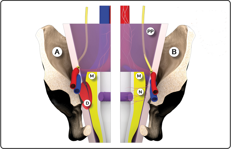
Three-dimensional model: Anatomic state before total mesorectal excision (TME) (A); risk of defect (D) in mesorectum (M) and parietal peritoneum (PP). Anatomic state after introduction of TME (B): intact mesorectum (M) and nerve-sparing resection (N) (Use VIPicture App).