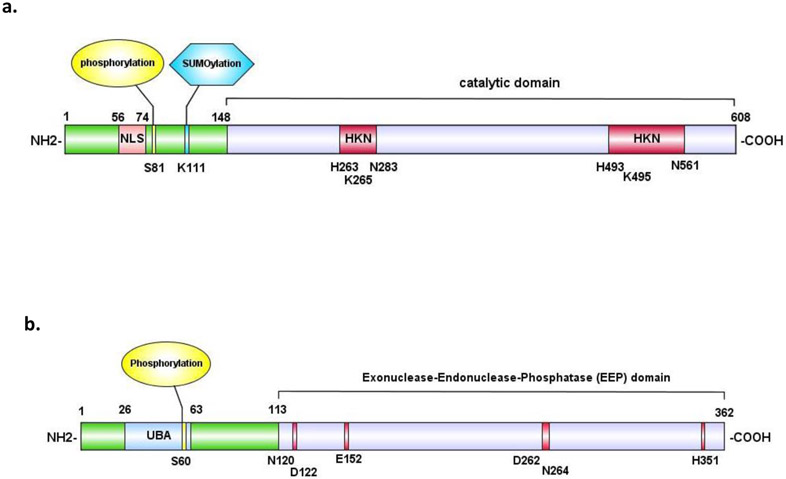
Figure 3. Domain structures of human tyrosyl-DNA phosphodiesterases.
a. domain structures of human TDP1. The nuclear localization signal (NLS) is highlighted in pink, the known phosphorylation site in yellow and the known SUMOylation site in blue. The phosphodiesterase motifs (HKN) are highlighted in magenta. b. domain structure of human TDP2. The ubiquitin-associated domain (UBA) is highlighted in cyan. The known phosphorylation site is highlighted in yellow. Catalytic sites are highlighted in magenta. The UBA (ubiquitin binding domain) in the N-terminus is colored in blue.

