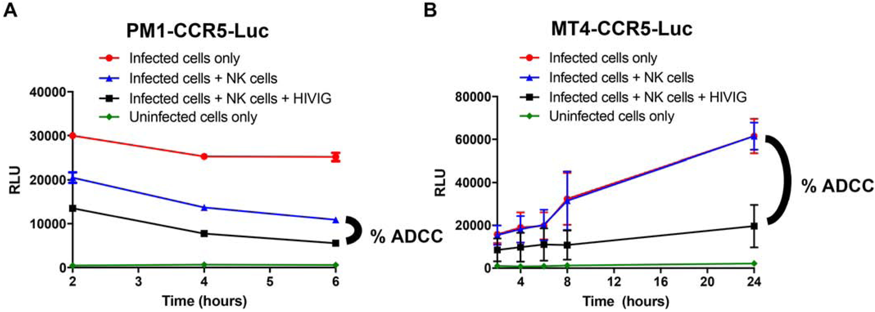
Figure 2. MT4-CCR5-Luc, but not PM1-CCR5-Luc, cells are resistant to CD16+KHYG-1 cell killing in the absence of antibodies.

Relative light units (y-axis) over time (x-axis) in NL43-infected PM1-CCR5-Luc (A) and MT4-CCR5-Luc (B) cells without antibody or NK cells (red circle), with NK cells but no antibody (blue triangle), and with NK cells and 500 μg/mL of HIVIG (black square). ADCC is determined as the percent difference as graphed. Uninfected cells (green diamond) represents background RLU reading. Data from 2 independent experiments with 3 replicates each. Bars shows standard error mean (SEM).
