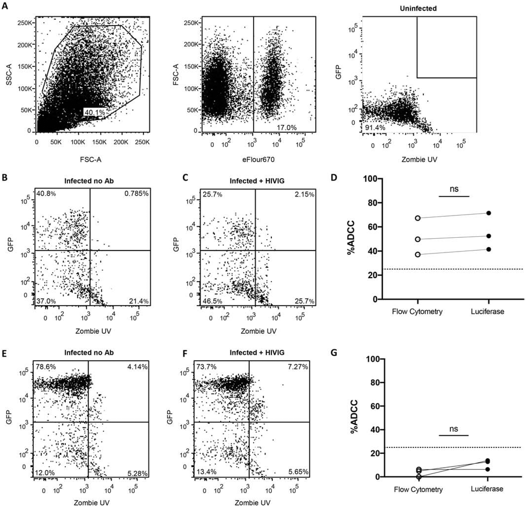
Figure 5. Proposed ADCC assay has been validated via flow cytometry.

MT4-CCR5-Luc cells were stained with eFluor670 prior to use in assay. (A) Gating strategy is shown for the condition with uninfected MT4-CCR5-Luc cells and NK cells, which represents background signal. The final panel shows live GFP+ target cells (Q1). (B) NL43-infected MT4-CCR5-Luc cells and NK cells (no antibody added). (C) NL43-infected MT4-CCR5-Luc cells, NK cells, and HIVIG. (D) Percent ADCC calculated using luciferase readout and percent ADCC calculated as the change in percentage of live GFP+ target cells (Q1) in the flow cytometry analysis (p = 0.25, Wilcoxon matched-pair test). (E) NL43-ΔEnv-Luc-VSVG-infected MT4-CCR5-Luc cells and NK cells (no antibody added). (F) NL43-ΔEnv-Luc-VSVG-infected MT4-CCR5-Luc cells, NK cells, and HIV Ig. (G) Nonspecific cytotoxicity of NL43-ΔEnv-Luc-VSVG-infected MT4-CCR5-Luc cells observed using luciferase and flow cytometry readout.
