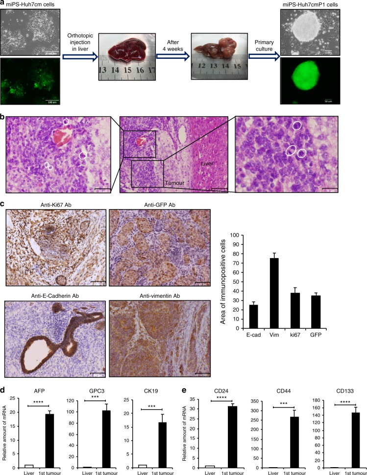
Fig. 1. Tumorigenicity of miPSCs treated with the conditioned medium of Huh7 cells.
a Representative scheme of the orthotopic injection of miPS-Huh7cm cells (miPS treated with Huh7cm for 4 weeks) into the liver and the formed tumour after 4 weeks of injection. Scale bars represent 100 and 50 µm. b Histopathological features of the primary tumours were evaluated by H&E staining, showing the presence of mitotic figures (white cycle). Scale bars represent 64, 32 and 16 µm. c Immunostaining of the malignant tumours for Anti-ki67 Ab, Anti-GFP Ab, Anti-E-Cadherin Ab and Anti-Vimentin Ab; and histogram showing area of immunopositive cells of each marker. All data were from three independent experiments (n = 3). Scale bars represent 64 μm. d RT-qPCR analysis for liver cancer-associated biomarkers in the derived tumour tissue compared to liver. All data were from three independent experiments (n = 3) (***p < 0.0001, ****p < 0.00001). e RT-qPCR analysis of CSC markers in the derived tumour tissue compared to liver. All data were from three independent experiments (n = 3) (***p < 0.0001, ****p < 0.00001).

