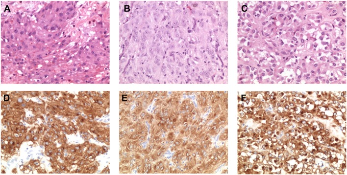
Figure 2.
Morphological and immunohistochemical analysis of low-allele-fraction KIT-mutant GIST. (A) GIST_260 appears as an epithelioid gastrointestinal stromal tumor comprising polymorphous cells arranged in nests and sheets with eosinophilic cytoplasm and peripherally placed nuclei, with mild nuclear atypia and no necrosis. The mitotic rate is 3/50 high power fields (HPF). (Hematoxylin and eosin stain: original magnification x 200). (B) GIST_241 is composed by spindle cell arranged in short fascicles and whorls, with pale eosinophilic fibrillary cytoplasm, ovoid nuclei, and ill-defined cell borders. The mitotic rate is 20/50 HPF. (Hematoxylin and eosin stain: original magnification x 200). (C) GIST_307 appears as an epithelioid gastrointestinal stromal tumor, composed by round cells with clear to eosinophilic cytoplasm arranged in sheets and nests, with low-grade nuclear atypia, mild nuclear pleomorphism and indistinct nucleoli. The mitotic rate is 2/50 HPF. (Hematoxylin and eosin stain: original magnification x 200). (D) CD117/KIT staining in GIST_260 reveals a strong positivity with membrane, cytoplasmic and “dot-like” Golgi staining (original magnification x 200). (E) KIT staining in GIST_241 shows a diffuse cytoplasmic staining (original magnification x 200). (F) Immunohistochemical analysis of CD117 expression in GIST_307 shows a strong and diffuse cytoplasmic and paranuclear Golgi-like staining (original magnification x 200).

