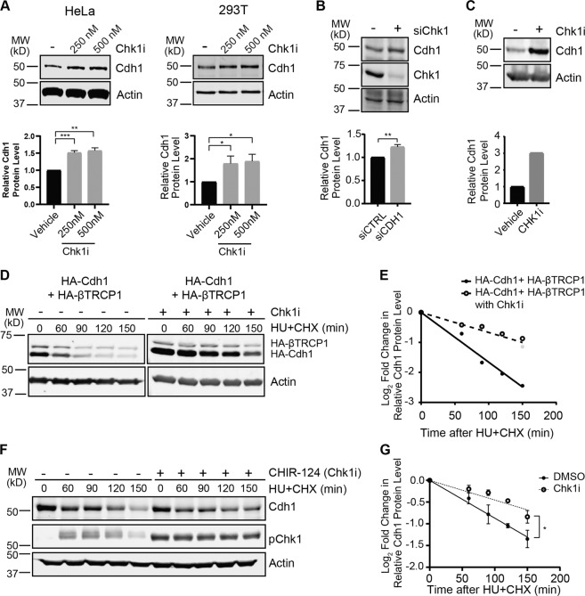
Fig. 1. Chk1 modulates Cdh1 stability.
a Inhibition of Chk1 increases the stability of endogenous Cdh1. Immunoblot analysis of whole-cell lysates derived from both HeLa and 293T cells treated with increasing doses of the Chk1 inhibitor (Chk1i), CHIR-124, for 5 h before harvesting. Lower panels, Cdh1 band intensities were normalized to actin and then further normalized to vehicle (n = 3). b Depletion of endogenous Chk1 leads to increased levels of endogenous Cdh1. Immunoblot analysis of whole-cell lysates derived from 293T cells transfected with the indicated siRNA (non-targeting siCTRL, −; siChk1, +). Lower panels, Cdh1 band intensities were normalized to actin and then further normalized to siCTRL transfected cells (n = 3). c Mitotic population does not contribute to the increased endogenous Cdh1 level after Chk1 inhibitor treatment. Immunoblot analysis of whole-cell lysates derived from HeLa cells after removal of the mitotic population via a mitotic shake-off post Chk1 inhibitor, CHIR-124 (500 nM) treatment for 5 h. Lower panel, Cdh1 band intensities were normalized to actin and then further normalized to vehicle-treated cells. d, e Inhibition of Chk1 increases the half-life of transfected Cdh1 in 293T cells. d Immunoblot analysis of whole-cell lysates derived from 293T cells, transfected with HA-Cdh1 and HA-βTRCP1 constructs. Cells were treated with both hydroxyurea (HU) and Chk1 inhibitor (Chk1i), CHIR-124 (500 nM), for another 4 h before addition of 50 µg/ml cycloheximide (CHX). At the indicated time points, whole-cell lysates were prepared for immunoblot analysis. e Quantification of the band intensities in (d). Cdh1 band intensities were normalized to actin and then further normalized to t = 0 controls. f, g Inhibition of Chk1 increases the half-life of endogenous Cdh1 in 293T cells. f. 293T cells were treated with both hydroxyurea (HU) and Chk1 inhibitor (Chk1i), CHIR-124 (500 nM), for another 4 h before addition of 50 µg/ml cycloheximide (CHX). At the indicated time points, whole-cell lysates were prepared for immunoblot analysis. g Quantification of the band intensities in (f) (n = 3). Mean and SEM are provided. Significance in panels (a), (b), and (g) was determined by a one-tailed unpaired T-test (*p < 0.05, **p < 0.005, and ***p < 0.0005).

