
Figure 7.
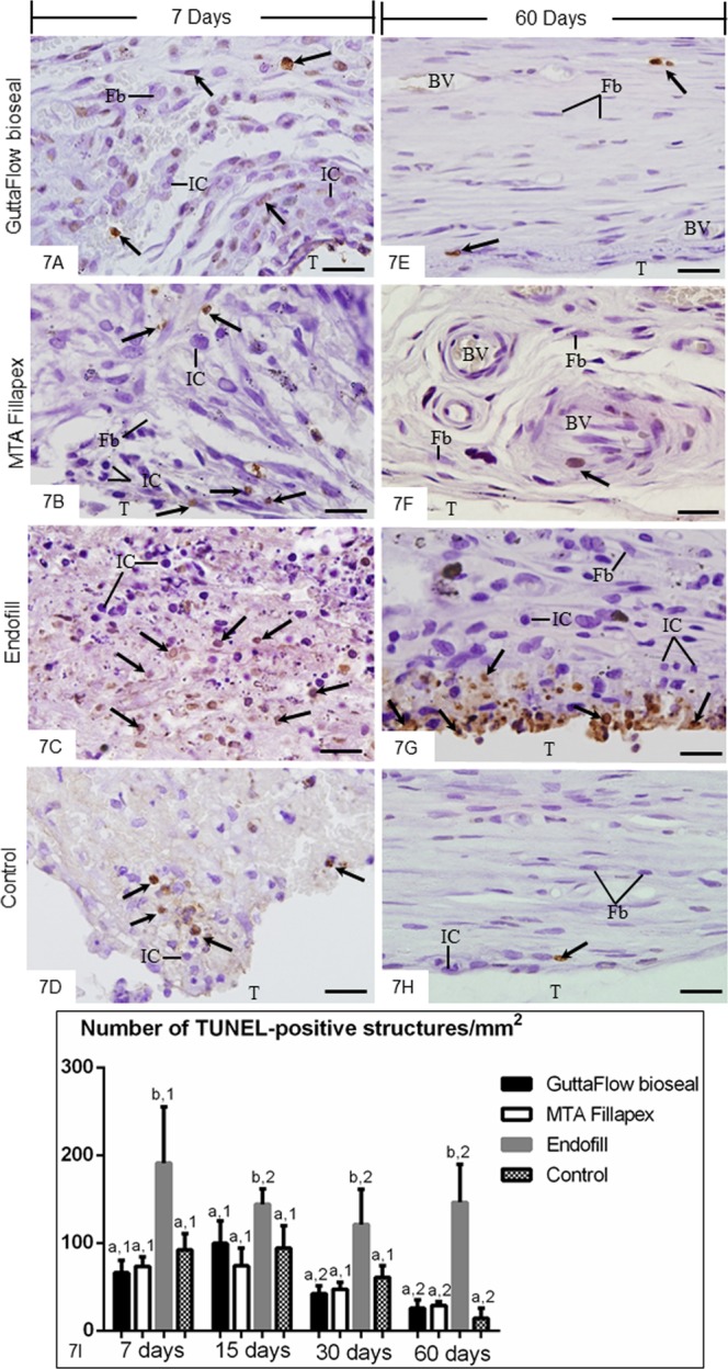
(A–H) Light micrographs showing portions of the capsules adjacent to the opening of the tubes implanted in the subcutaneous for 7 (A–D) and 60 (E–H) days. The sections were subjected to the TUNEL method and counterstained with haematoxylin. Round/ovoid TUNEL-positive structures (arrows) are observed among inflammatory cells (IC) and fibroblasts (Fb). An accentuated number of TUNEL-positive structures (arrows) is observed in the capsules of EF (C,G). T, space of the tube implanted; BV, blood vessel. Bars: 20 µm. (I) Graphic showing the number of TUNEL-positive structures per mm2 (expressed as mean ± standard deviation) in the capsules of GFB, MTAF, EF and CG groups at 7, 15, 30 and 60 days. The comparison among groups in the same period is indicated by superscript letters; same letters = no statistically significant difference. The analysis of each group over time is indicated by superscript number; same numbers = no statistically significant difference. N = 5 specimens per group at each time point. Tukey’s test (p ≤ 0.05).