
Figure 8.
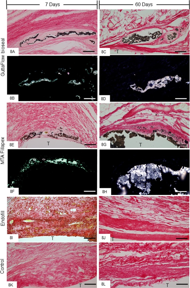
(A–L) Light micrographs showing portions of the capsules adjacent to the opening of the tubes implanted in the subcutaneous for 7 and 60 days. (A,C,E,G,I–L) The sections were subjected to von Kossa histochemical method and counterstained with picrosirius. von Kossa-positive structures (black colour) are seen dispersed by capsules of GFB (A,C) and MTAF (E,G) groups. von Kossa-positive structures are not observed in the EF (I,J) and CG groups (K,L). (B,D,F,H) Light micrographs showing unstained sections analyzed under polarized light. Note birefringent structures in the corresponding regions of the capsules shown in the sections submitted to the von Kossa method. T, space of the tube implanted; Bars: 65 µm.