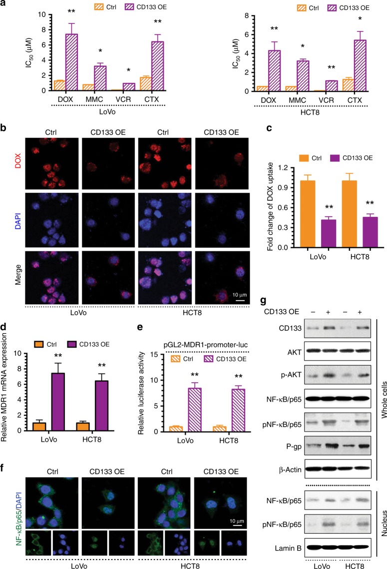
Fig. 3. CD133 promotes MDR via the AKT/NF-κB/MDR1 signalling in CRC.
a The IC50 values of DOX, MMC, VCR and CTX in LoVo, HCT8 and their CD133 overexpressing (CD133 OE) cells were determined with a CCK-8 assay. b Intracellular distribution of DOX (red) at the 6th h after 1 h incubation with 1 μg/ml DOX. c Quantitative DOX profiles over 6 h. d qPCR showing MDR1 mRNA expression. e Luciferase reporter assay showing MDR1 gene promoter activity. f Immunofluorescence showing the localisation of NF-κB/p65. g Western blots showing protein profiles of CD133, total AKT, p-AKT, total NF-κB/p65 (whole cells or nucleus), p-NF-κB/p65 (whole cells or nucleus) and P-gp. *p < 0.05, **p < 0.01. Each bar represents the mean ± SD of three independent experiments.

