Figure 3.
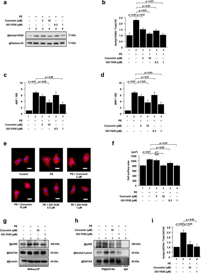
GO-Y030 at a dose 1/10th that of curcumin significantly suppressed PE-induced hypertrophic responses in cardiomyocytes. Primary cultured cardiomyocytes were treated with 3 or 10 μM curcumin, or with 0.3 or 1 μM GO-Y030 and were stimulated with 30 μM PE. (a) Histone fractions isolated from these cells were subjected to western blotting using anti-acetyl-histone H3 (Lys9) antibodies and anti-histone H3 antibodies. Full-length blots are presented in Supplementary Fig. S6. (b) The levels of acetylated histone H3K9 and total histone H3 were quantified. The data are presented as the mean ± SEM of three individual experiments. (c,d) Total RNA was extracted from the cells, and quantitative PCR was performed for ANF (c), BNP (d), and 18S. The data are presented as the mean ± SEM of three individual experiments. (e) Immunofluorescence staining was performed using anti-MHC antibodies and Alexa Fluor 555-conjugated anti-mouse IgG. Scale bar: 20 μm. (f) The surface area of these cells was measured using ImageJ software. All data are presented as the mean ± SEM of three individual experiments. (g) Nuclear extracts prepared from primary cultured cardiomyocytes were subjected to western blotting with anti-p300 antibodies, anti-GATA4 antibodies, and anti-β-actin antibodies. Full-length blots are presented in Supplementary Fig. S7. (h) The nuclear extracts were immunoprecipitated with goat anti-GATA4 polyclonal antibodies and subjected to western blotting with anti-p300 antibodies, anti-acetyl-lysine antibodies, and anti-GATA4 antibodies. The original images are presented in Supplementary Fig. S8. (i) The levels of acetylated GATA4 were quantified. The data are presented as the mean ± SEM of three individual experiments.

