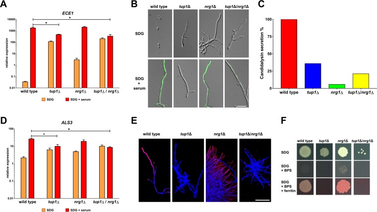
FIG 1.
Tup1 is required for high-level expression of ECE1 and ALS3. (A) Total RNA was isolated from the indicated strains after 6 h of growth in SDG with or without 10% human serum. ECE1 transcription was normalized against ACT1 and the control RNA (wild type, 5 h YPD, 37°C). Asterisks indicate significant transcription differences in a mutant compared to the wild type after growth in SDG with serum (P ≤ 0.05, two-tailed, unpaired Student's t test). (B) The indicated strains with integrated pECE1-GFP cassettes were grown for 6 h at 37°C in SDG with or without serum prior to microscopy. Shown are the overlays of the DIC channel and the GFP channel. Scale bar, 20 μm. (C) Candidalysin secretion was measured by LC-MS/MS after 18 h of growth in YNBS (pH 7.2). Candidalysin contents measured for wild-type hyphae were defined as 100%. (D) The total RNA isolated as described for panel A was used to determine the normalized relative expression levels of ALS3. Asterisks indicate significant transcription differences in a mutant compared to the wild type after growth in SDG with serum (P ≤ 0.05, two-tailed, unpaired Student's t test). (E) After 6 h of growth in SDG with 10% human serum at 37°C, cells of the indicated strains were stained first with a monoclonal anti-Als3 antibody (pink signal) and then with calcofluor white (blue signal). Overlays of the images taken in the Cy5 and DAPI (4′,6-diamidino-2-phenylindole) channel are shown. Scale bar, 50 μm. (F) From an overnight culture, 103 cells of the indicated strains were dropped on SDG medium with or without an iron chelator (bathophenanthroline disulfonate [BPS]) and with or without ferritin. The plates were grown for 3 days at 37°C in 5% CO2 before images were taken.

