FIG 5.
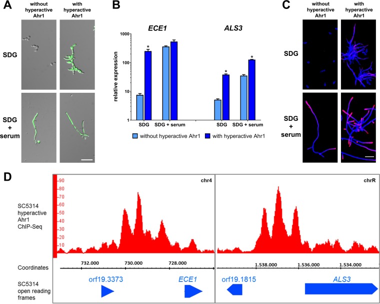
Hyperactive Ahr1 induces high-level expression of ALS3 and ECE1. (A) The indicated strains with or without the hyperactive Ahr1 were grown in SDG or SDG with 10% human serum. Pictures were taken after 6 h of growth at 37°C. Shown are the overlays of the DIC and the GFP channels. Scale bar, 20 μm. (B) After 6 h of growth, total RNA of the strains grown as described for panel A was isolated and used for determination of relative gene expression levels. Asterisks indicate significant changes (P ≤ 0.05, two-tailed, unpaired Student's t test) in mutants with hyperactive Ahr1 compared to their background strains without the hyperactive allele. (C) Cells of the indicated strains were grown for 6 h in SDG with 10% human serum at 37°C and were then stained with a monoclonal anti-Als3 antibody (pink signal), followed by a second staining with calcofluor white (blue signal). Shown are the overlays of the images taken in the Cy5 and DAPI channels. Scale bar, 20 μm. (D) ChIP-Seq shows direct binding of hyperactive Ahr1 to the promoters of ECE1 and ALS3. Genomic DNA used for ChIP-Seq was isolated from the wild-type strain with hyperactive Ahr1 after 6 h growth in SDG medium at 37°C. The binding peaks as shown in the IGB viewer are displayed.

