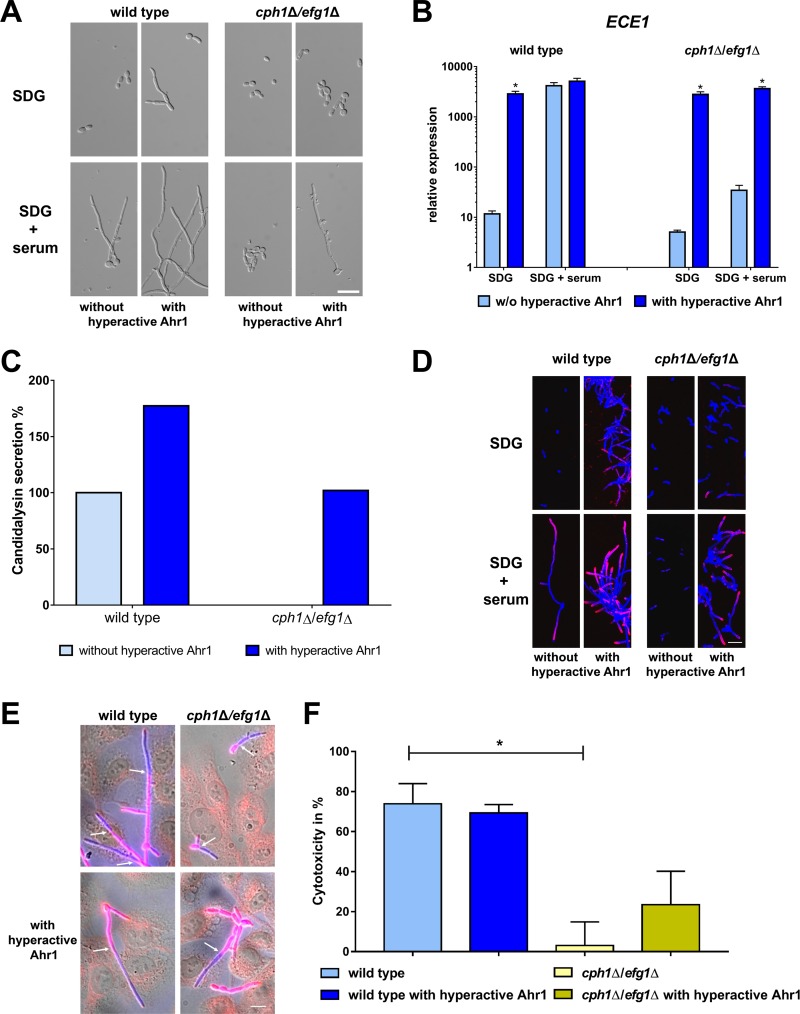
FIG 7.
Hyperactive Ahr1 activates ECE1 and ALS3 expression in the nonfilamentous cph1Δ/efg1Δ mutant. (A) Wild type SC5314 and cph1Δ/efg1Δ mutant with or without the hyperactive Ahr1 were grown for 6 h at 37°C in either SDG or SDG with 10% human serum prior to microscopy. Scale bar, 20 μm. (B) After 6 h growth under the same conditions as described for panel A, total RNA of these strains was isolated and used for determination of relative ECE1 expression levels. Asterisks indicate significant changes (P ≤ 0.05, two-tailed, unpaired Student's t test) in mutants with hyperactive Ahr1 compared to their background strains without the hyperactive allele. (C) Candidalysin secretion of wild-type and cph1Δ/efg1Δ strains with or without the hyperactive Ahr1 was measured by LC-MS/MS after 18 h growth in YNBS (pH 7.2). Candidalysin secreted by wild-type hyphae was defined as 100%. (D) Cells of the indicated strains were grown for 6 h in SDG with 10% human serum at 37°C and then stained with the Als3 antibody (pink) and calcofluor white (blue signal). Shown are the overlays of the images taken in the Cy5 and DAPI channels. Scale bar, 20 μm. (E) TR-146 oral epithelial cells infected with wild-type and cph1Δ/efg1Δ strains with or without hyperactive Ahr1 after 4 h coincubation. C. albicans outside human cells is shown in pink; C. albicans inside human cells is shown in blue. Arrows mark the point of invasion. Scale bar, 20 μm. (F) Cytotoxicity of the indicated strains was determined by the release of LDH from infected TR-146 cells after a 24 h coincubation. Asterisks mark significant changes (P ≤ 0.05, two-tailed, unpaired Student's t test) to values of the wild-type strain.

