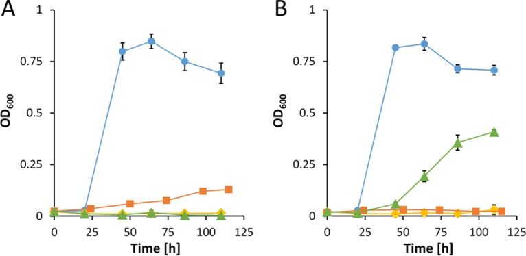
FIG 5.

Growth of strains ΔpedE ΔpedH (blue circles), ΔpedE ΔpedH ΔglpFKRD (yellow diamonds), ΔpedE/H Δglp-Tn7M-pedE (orange squares), and ΔpedE/H Δglp-Tn7M-pedH (green triangles) in M9 minimal medium supplemented with 20 mM glycerol in the absence (A) or presence (B) of 10 μM La3+. Incubation was performed in 96-well microtiter plates in a rotary shaker (Forma; Thermo Scientific) at 28°C and 220 rpm. Data represent averages of results from biological triplicates with corresponding standard deviations.
