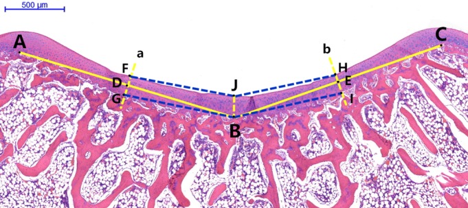
Figure 3.

Axial microscopic view of trochlear groove. A and C are highest points of the medial and lateral condyles of the femoral trochlea. B is the deepest point of the bony trochlear groove. D and E are the midpoints of lines AB and BC, respectively. Line a passes through point D perpendicular to line AB. FG is the cartilage thickness of the medial facet. Line b passes through point E perpendicular to line CB. HI is the cartilage thickness of the lateral facet. The distance JB is the thickness of the central cartilage (deepest sulcus position). Angle FJH is the cartilaginous sulcus angle. Angle GBI is the bony sulcus angle. F and H are the points at which the cartilaginous trochlear groove intersects with lines a and b, respectively. G and I are the points at which the bony trochlear groove intersects with lines a and b, respectively.
