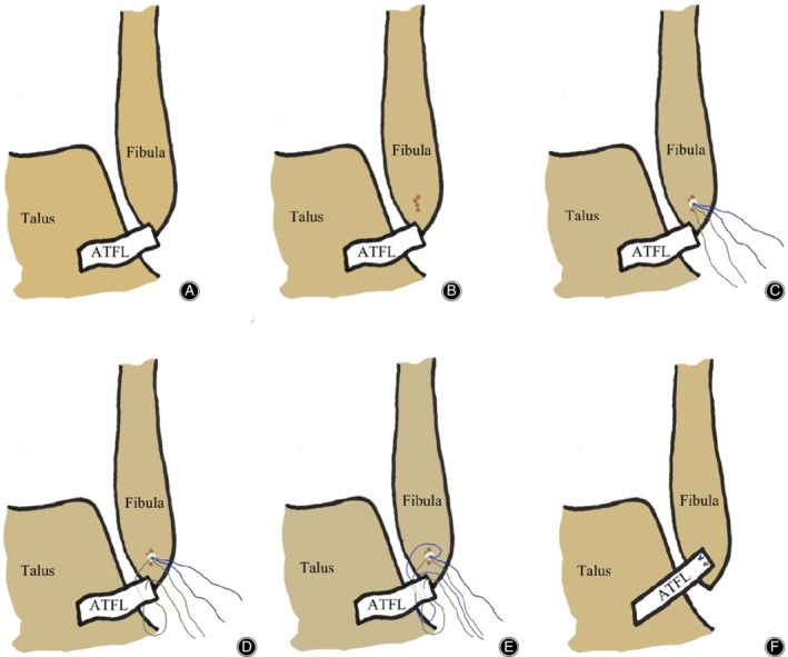
Figure 1.

Surgical diagrams of arthroscopic anatomical repair of anterior talofibular ligament (ATFL). (A) The intact ATFL was avulsed from the fibula. (B) The bone of the fibula footprint was freshened using a Pituitary Rongeur or 1.0 mm Kirschner wire drill. (C) A double wire anchor with a diameter of 3.5 mm was inserted in the middle area of the fibula footprint. (D) The suture method of the first anchor sutural wire. (E) The suture method of the second anchor sutural wire. (F) The ATFL was sutured.
