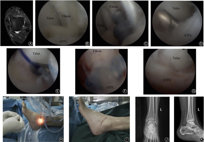
Figure 2.

A 32‐year‐old male with recurrent sprain of the left ankle with unstable walking for 18 months. The symptoms were not relieved after 12 months of regular conservative treatment. Arthroscopic anatomical repair of ATFL was performed. (A) Preoperative MRI plain scan of the left ankle suggested that continuity of ATFL was interrupted. (B) Arthroscopic exploration showed horizontal avulsion of ATFL from the fibular stop point and relaxation of ATFL without tension. (C) The footprint region of the fibula was observed under arthroscopy. After freshening, a double wire anchor with a diameter of 3.5 mm was inserted. (D) The ATFL was sutured with a suture hook under arthroscopy. (E) Under arthroscopy, the anchor sutural line was guided through the ATFL after the PDS sutural line was used to pass through the suture hook. (F) Fibular side of the ATFL was sutured with a knot pusher. (G) The ATFL returned to normal tension and strength after suture under arthroscopy. (H) During the operation, the PDS sutural line was used to guide the anchor wire through the external phase. (I) The surgical approach after suture. (J, K) Anterior–posterior and lateral X‐ray films of the ankle after surgery.
