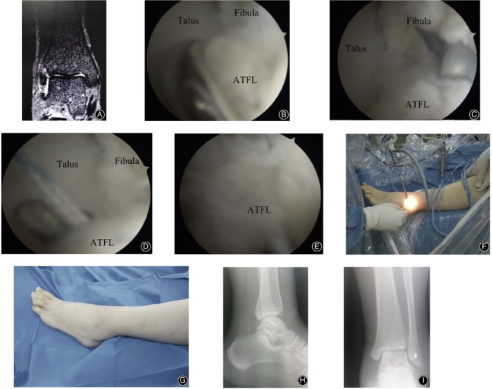
Figure 3.

A 28‐year‐old female. The symptoms of CLAI were still presented after 8 months of conservative treatment. (A) Preoperative coronal MRI scan of the left ankle suggested the interruption of ATFL. (B) Flaccid and tension‐free ATFL that avulsed from the fibula point was observed under the arthroscopy. (C) A double wire anchor with a diameter of 3.5 mm was inserted into the fibular footprint region. (D) The ATFL was sutured with a suture hook under arthroscopy. (E) The ATFL was sutured and it returned to normal tension under arthroscopy. (F) External image of the arthroscopic procedure during the operation. (G) The portals of the surgery after the operation. (H) Lateral X‐ray of the ankle after surgery. (I) Anterior–posterior X‐ray of the ankle after surgery.
