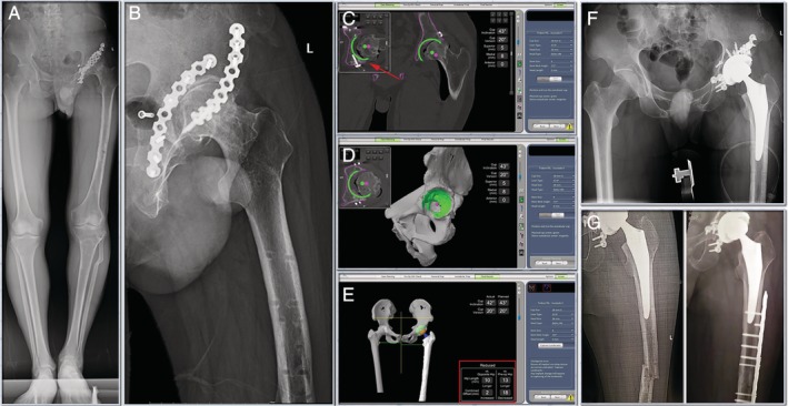
Figure 2.

(A) & (B) Preoperative radiographs. The patient suffered from post‐traumatic arthritis of the left hip, with internal fixation on the anterior and posterior walls of the acetabulum. (C) & (D) CT based pre‐operative planning allowed to plan the cup away from the existing hardware (red arrow) in pelvis. (E) & (F) Robotic arm was used to execute that plan. And the leg length discrepancy was 10mm (red square). The simulation results of Mako system were the same as those of actual post‐operative X‐ray. (G) Patient's previous fracture site on femur recurred 1 month later, and then the fracture was held together by plate and screws.
