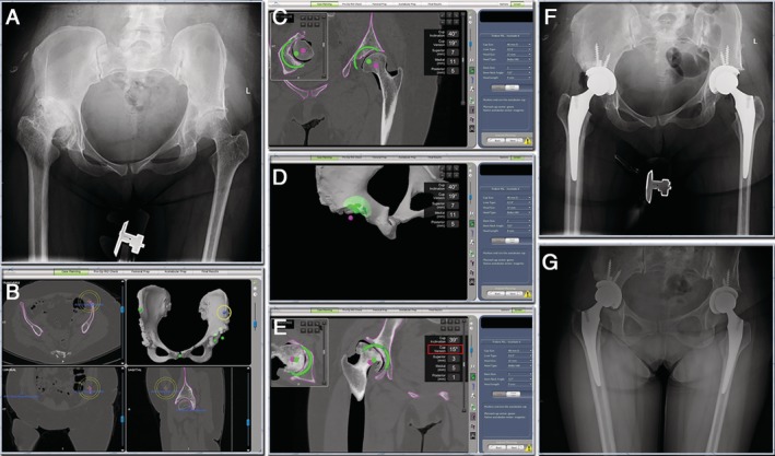
Figure 3.

(A) Pre‐operative radiographs. The patient suffered from bilateral DDH. (B) & (C) & (D) & (E) CT based pre‐operative planning. Left ASIS was absent due to previous osteotomy (yellow circle). The position of the left ASIS reference point was carefully adjusted to make the pelvis axis as horizontal as possible. The cups were planned to reconstruct with the high hip center technique. The right cup anteversion was planned to be lower than usual (red square). (F) The post‐operative X‐rays showed that the robotic arm assisted THA had achieved the surgery plan. (G) Radiograph 3‐months post‐operative showed no change in the position of the prosthesis.
