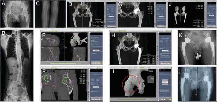
Figure 4.

(A) & (B) & (C) Pre‐operative radiographs. Bilateral hips and knees have fused because of ankylosing spondylitis. (D) & (E) & (F) & (G) & (H) CT based pre‐op planning. The 3 acetabular align points that should had been set at the acetabulum were set at the great trochanter and acetabular rim (yellow circle). (I) & (J) & (K) Robotic arm was used to execute that plan. In the registration step of the operation, the circumference of the joint (great trochanter, femoral neck, acetabulum rim) was registered instead of the acetabulum (red circle). The robotic arm assisted THA had achieved the surgery plan. (l) Radiograph 3‐months post‐operative showed no change in the position of the prosthesis.
