FIG 4.
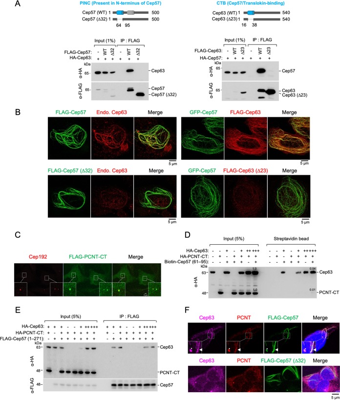
Mutual requirement of Cep57 PINC and Cep63 CTB for proper Cep57-Cep63 interaction and their colocalization. (A) Schematic diagram showing the Cep57(Δ32) and Cep63(Δ23) mutants lacking residues 64 to 95 (i.e., the PINC motif) of Cep57 and lacking residues 16 to 38 (i.e., the CTB motif) of Cep63, respectively. Gray bars indicate α-helices predicted by the PSIPRED server. (B) 3D-SIM images of U2OS cells transfected with the indicated constructs and immunostained with either anti-Cep63 antibody (left) or anti-FLAG antibody (right). Endo., endogenous. (C) Confocal analysis was performed with U2OS cells transfected with FLAG–PCNT-CT. The resulting cells were immunostained with anti-FLAG and anti-Cep192 antibodies. Boxes indicate areas of image enlargement. (D) Pulldown assays of biotinylated Cep57(61–95) with HEK293 lysates expressing HA-Cep63 and HA–PCNT-CT. Streptavidin-agarose-immobilized Cep57(61–95) and its coprecipitates were analyzed by anti-HA immunoblotting. Note that while Cep57(61–95) efficiently coprecipitates Cep63, it fails to significantly coprecipitate PCNT-CT containing the PACT domain, a suggested Cep57 PINC-binding region (18). (E) IP and immunoblot analyses using HEK293T cells cotransfected with the indicated constructs. Note that a Cep57 N-terminal construct containing the entire centrosomal localization domain (24) also preferentially binds to Cep63 but not PCNT-CT. (F) Confocal images of U2OS cells transfected with either FLAG-Cep57 WT or FLAG-Cep57(Δ32) and immunostained with either anti-PCNT, Alexa Fluor 647-conjugated anti-Cep63, or anti-FLAG antibodies. Nuclear DNA was labeled with DAPI (blue). Boxes indicate areas of enlargement. Asterisks indicate centrosomes. Arrowheads indicate endogenous Cep63 and PCNT recruited to a FLAG-Cep57 cable.

