Figure 2.
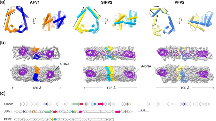
Comparison of the filamentous viruses AFV1, SIRV2 and PFV2. (a) MCP dimer (asymmetric unit) comparison of AFV1, SIRV2 and PFV2. The MCP1 of AFV1, SIRV2 and PFV2 are colored in orange, gold and yellow, respectively. The MCP2 of AFV1, SIRV2 and PFV2 are colored in blue, cyan and light blue, respectively. The N-terminal helices of MCP1 in AFV1, SIRV2 and PFV2 are marked with black arrows. (b) Wrapping of A-DNA in AFV1, SIRV2 and PFV2. Five MCP dimers are displayed: one MCP dimer is colored as in (a); the other four colored in gray. Proteins are shown in ribbon representation (top) and as surfaces (bottom). (c) Comparison of genome maps of rudivirus SIRV2 (NC_004086), lipothrixvirus AFV1 (NC_005830) and tristromavirus PFV2 (MN876844). Homologous genes (E < 1e–04) are indicated with the same colors. The homology between the MCPs of PFV2 (yellow and cyan ORFs) and those of the other two viruses is not recognizable by sequence similarity searches.

