Figure 2.

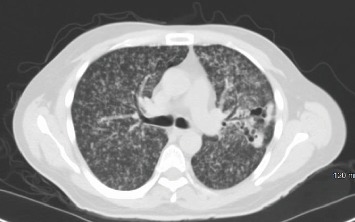
Thoracic CT scan without contrast demonstrating numerous, bilateral tiny nodular densities, most prominently in the upper lobes and confirmed a lingular cavitary lesion.

Thoracic CT scan without contrast demonstrating numerous, bilateral tiny nodular densities, most prominently in the upper lobes and confirmed a lingular cavitary lesion.