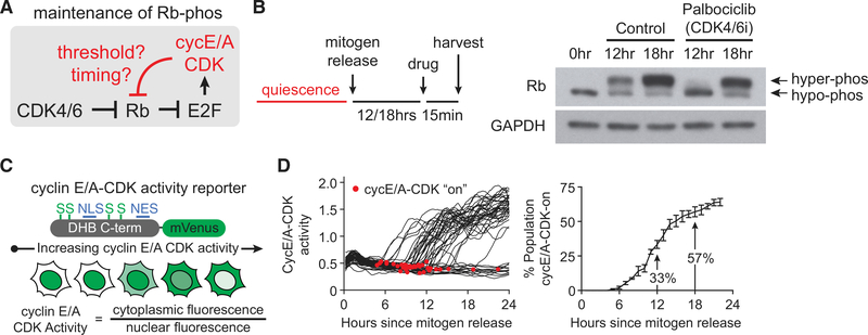
Figure 1. Heterogeneity in Cyclin E/A-CDK Activity Obscures Regulatory Mechanisms of Rb Hyperphosphorylation in Bulk-Cell Analysis.
(A) Schematic highlighting possible threshold or cell-cycle timing-dependent maintenance of Rb hyperphosphorylation by cyclin E/A-CDK following acute CDK4/6 inhibition.
(B) MCF-10A cells mitogen-released for indicated times prior to harvesting and western blot analysis. For 12 and 18 h time points, cells were treated with palbociclib 1 μM or vehicle (0.1% DMSO) for 15 min prior to harvesting.
(C) Schematic of cyclin E/A-CDK activity reporter.
(D) MCF-10A expressing cyclin E/A-CDK activity reporter and H2B-mTurquoise (for tracking) were mitogen-released and live-cell-imaged. Left: 50 random traces with automated detection of initial activation shown for illustration. Right: percentage of cells that have started to activate cyclin E/A-CDK by a given time since mitogen release. Error bars are SD; n = 3 replicates; n ≥ 105 cells per replicate.

