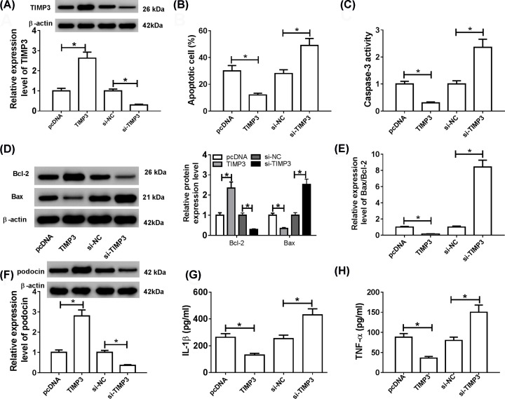
Figure 4. TIMP3 inhibited apoptosis and inflammation in HG-treated podocytes.
HG-stimulated podocytes were transfected with pcDNA, TIMP3, si-NC or si-TIMP3, respectively. (A) The protein level of TIMP3 was examined using Western blot. (B) Cell apoptotic rate was estimated by flow cytometry. (C–E) Caspase-3 activity, Bcl-2 and Bax levels, and Bax/Bcl-2 ratio were detected using Western blot analysis. (F) The expression of podocin was examined by Western blot assay. (G and H) The levels of inflammatory cytokines (IL-1β and TNF-α) were measured by ELISA analysis; *P<0.05.

