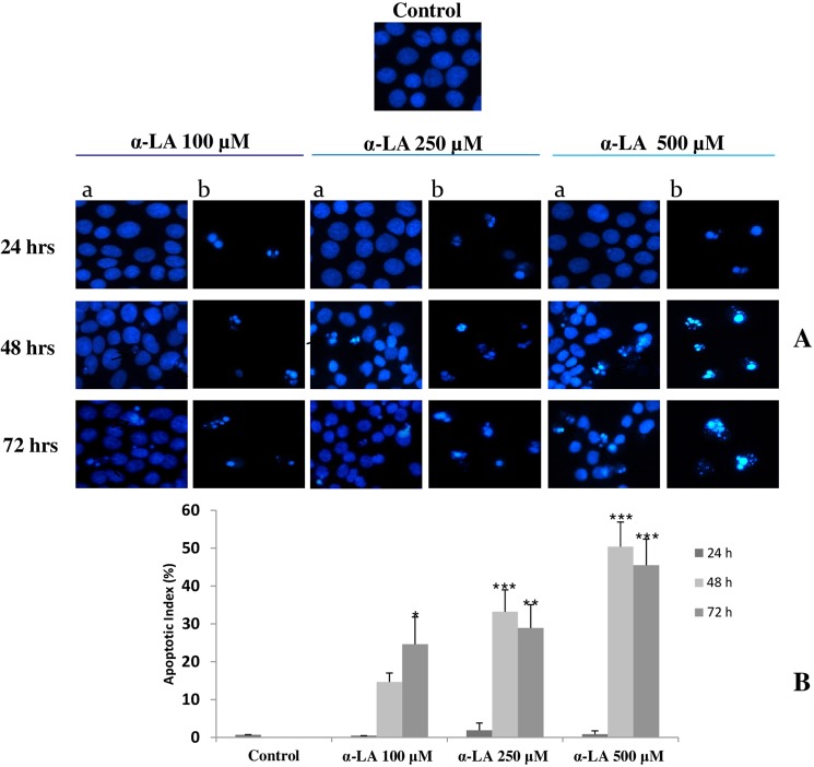
Figure 1.
Induction of apoptosis after exposure to increasing concentrations of α-LA (A) FaO cells were exposed to 100, 250 and 500 µM α-LA for 24, 48 and 72 hours and subjected to Hoechst 33258 nuclear staining; with a are indicated stained attached cells and with b apoptotic detached cells. (B) Percentage of apoptotic cells (Apoptotic Index). Data are shown as means ± S.E. Assay was performed in quadruplicate. Significantly different from control cells for p < 0.05*; p < 0.01 **; p < 0.001***.

