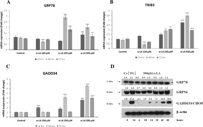
Figure 4.
Involvement of ER stress in the onset of apoptosis α-LA-mediated in FaO hepatoma cells. (A–C) Expression profiles of ER stress representative genes Bip/Grp78, Trib3 and Gadd34 were validated with Real Time PCR analysis using RNA isolated from three biological replicate. FaO cells were exposed to 100, 250 and 500 µM α-LA for 24, 48 and 72 hours. Results are expressed as means ± SE. Significantly different from control cells for p < 0.05*; p < 0.01 **; p < 0.001***. (D) Western blot analysis of GRP78, GRP94 and GADD153/CHOP in rat FaO hepatoma cells treated with 500 µM α-LA from 6 up to 48 hours. Cells treated with 0,5 μM Thapsigargin (TG) were used as positive control. β-actin was used as loading control. For densitometric analysis specific protein expression was normalized to β-actin expression and values (reported over each band) have been expressed as fold change respect to control. Each lane represents a pool of three individual samples. Western Blot images (D) have been cropped for clarity with full blot presented in Supplementary Fig. 6.

