Figure 7.
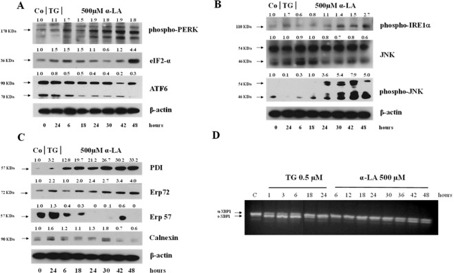
Analysis of PERK, IRE1 and ATF6 pathways and PDI proteins involved in UPR activation. Western blot analysis of (A) phospho-PERK, eIF2-α and ATF6, (B) phospho-IRE1α, total JNK and phospho-JNK (C) PDI, Erp72, Erp57 and calnexin in rat FaO hepatoma cells treated with 500 µM α-LA from 6 up to 48 hours. β-actin was used as loading control. For densitometric analysis specific protein expression was normalized to β-actin expression and values (reported over each band) have been expressed as fold change respect to control. Each lane represents a pool of three individual samples. TG: Thapsigargin. (D) Splicing of XBP-1 in rat FaO hepatoma cells after exposure to 0,5 uM TG from 1 up to 24 hours or 500 μM α-LA from 3 up to 48 hours. The data are representative of three different individual experiments. Images (A–D) have been cropped for clarity with full blot presented in Supplementary Fig. 7.

