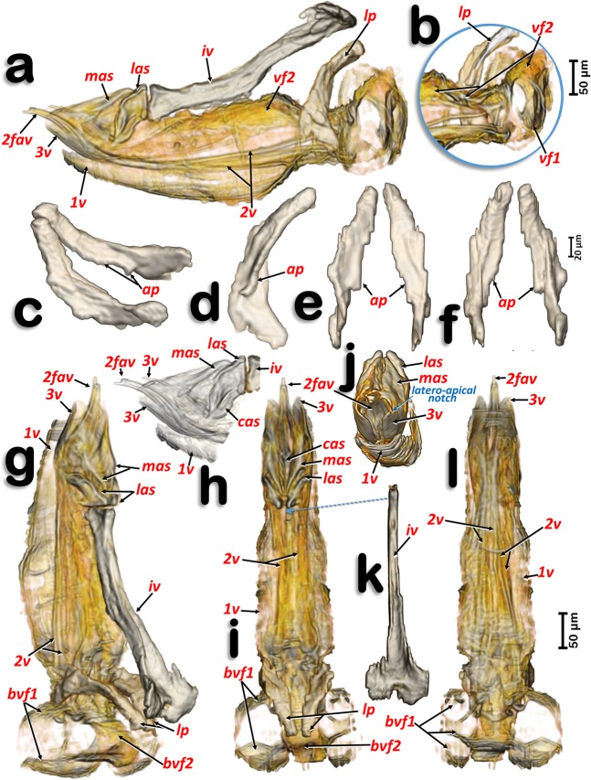
Figure 7.
Volume-rendered images of the ovipositor in different perspective views. Right-lateral (a,b,h), right dorso-lateral (g), dorsal (i), posterior (j) and ventral (l). Latero-basal plate sclerites in different views: right dorso-lateral (c), internal side of the left sclerite (d), posterior (e) and frontal (f). Detail of a right apical view, using software to show the apical sclerites and valvulae (h). Dorsal view of the intervalvular basal sclerite (k). Abbreviations: 1 v, 2 v and 3 v = 1st, 2nd and 3rd valvulae; 2fav = fused apical part of the 2nd valvula; ap = latero-basal plate internal apodeme; cas = centro-apical sclerite; iv = basal intervalvular sclerite; las = latero-apical sclerite; lp = latero-basal plate sclerite; mas = medio-apical sclerite; vf1 and vf2 = 1st and 2nd valvifera.

