Fig. 1.
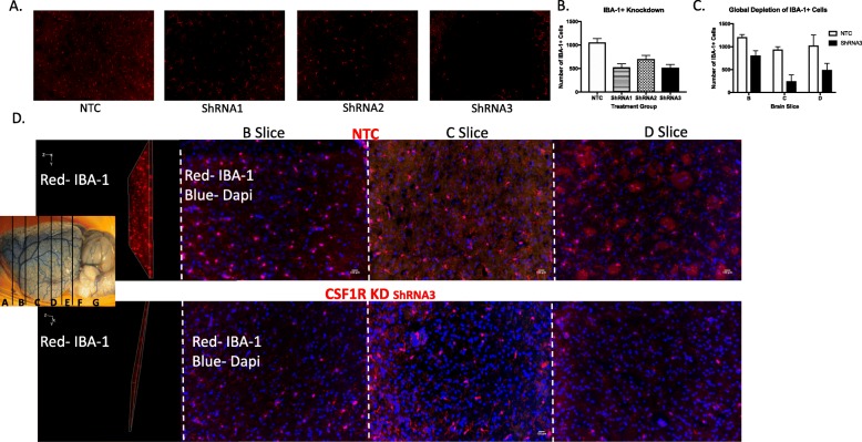
Bilateral CSF1R ShRNA injection resulted in a global knockdown of IBA-1+ cells brain 14 days post-injection. Images were derived from control animals and were Z stacked and quantified throughout the layer. Thirty-micrometer-thick sections were taken from the B, C, and D slice, both the layer and a representative image indicating the slides are illustrated in a and quantified in b. Three ShRNA constructs all targeted at the CSF1R were evaluated 14 days post-injection. Based on this evaluation, we choose the ShRNA with the best knockdown percentage to proceed with the study. Representative images of the chosen ShRNA and the quantification of the degree of knockdown are shown in C and D. ShRNA injection resulted in a reduction of IBA-1+ cells in the "slice" (b"d"), C slice ("d"c), and D slice ("d"d) (N = 4/group)

