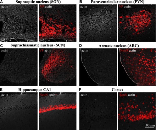
Figure 1.
Examination of actin organization in different areas of the rat brain. Confocal imaging of immunostaining for β-actin (white) and VP (red, A–C) or neuronal marker NeuN (red, D–F) analyzed in adult rat brain sections containing SON (A), PVN (B), SCN (C), ARC (D), hippocampus CA1 (E), cortex (CX; F), and other brain areas (Extended Data Fig. 1-1).

