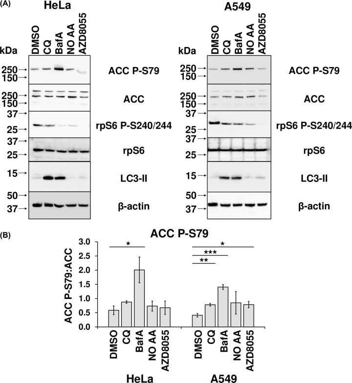
Figure 9. BafA enhances AMPK activity.
(A) Extracts were prepared from HeLa and A549 cells that had been treated with 1:1000 DMSO (vehicle), 50 µM CQ, 200 nM BafA, or 1 µM AZD8055 for 24 h. Alternatively, they were treated with serum-free DMEM for 24 h or for 23.5 h, followed by a 30-min treatment with Krebs–Ringer Bicarbonate buffer 20 mM glucose, but no added amino acids (NO AA). Cell lysates were analysed via immunoblotting with the indicated antibodies. Results are representative of three independent experiments. (B) ACC P-79 (relative to total ACC) was quantified by densitometric analysis, and represented as the mean of three independent experiments. Error bars represent ± standard error of the mean of the indicated number of independent experiments performed. Statistical significance was determined using unpaired Student’s t-test. Note: *P≤0.05, **P≤0.01, ***P≤0.001.

