
Figure 2: Histopathology, Fluorescence In Situ Hybridization and Immunohistochemistry (Case 1 & 4).
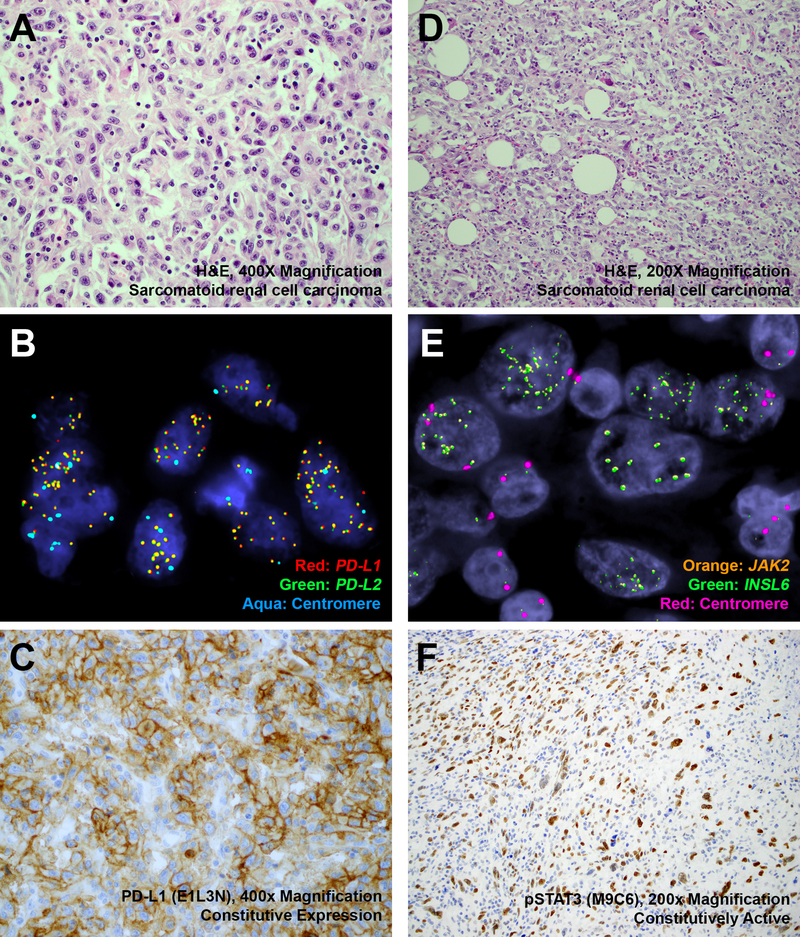
Representative H&E-stained images of 9p24.1-amplified sarcomatoid renal cell carcinomas ((a) Case 4, 2.7-fold amplification by MSK-IMPACT, × 400 magnification; (d) Case 1, 3.5-fold amplification by MSK-IMPACT, × 200 magnification), corresponding fluorescence in situ hybridization ((b) Red: PD-L1, Green: PD-L2, Aqua: Centromere; (e) Orange: JAK2, Green: INSL6, Red: Centromere) and immunohistochemistry ((c) PD-L1, × 400 magnification; (f) phospho-STAT3, × 200 magnification) have been depicted. MSK-IMPACT, Memorial Sloan Kettering Cancer Center Integrated Mutation Profiling of Actionable Cancer Targets.