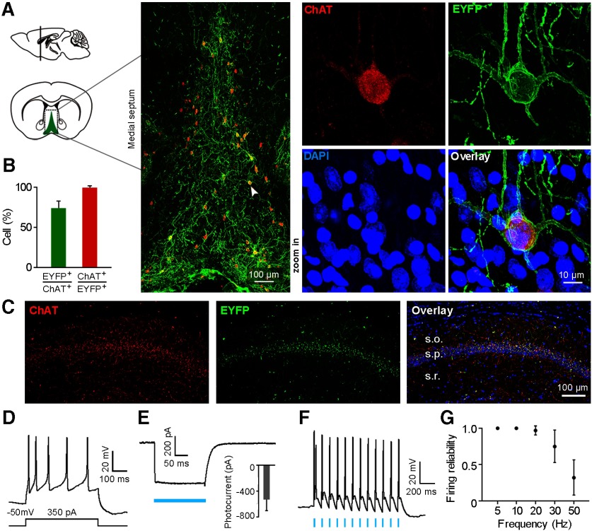
Figure 1.
ChR2 is strongly expressed in MS cholinergic neurons and mediates robust optogenetic activation in ChAT-ChR2-eYFP transgenic mice. A, Left, Schematic drawing shows MS in a coronal plane. Middle, Confocal image of boxed area on the left shows that ChR2-eYFP (green) is strongly expressed in ChAT-positive neurons (red). The neuron indicated by the arrowhead is shown at right in the high-magnification view. B, Percentages of ChAT neurons that are ChR2-eYFP positive, and vice versa (mean ± SD; n = 5 mice). C, Confocal image of a hippocampal coronal section shows cholinergic terminals colocalized with ChR2-EYFP in the hippocampal CA1 area, with the most abundant innervations found in stratum pyramidale. D, Action potentials induced by current injection in a ChR2-EYFP-expressing neuron from a brain slice patch-clamp recording. E, Blue light-evoked photocurrent in a ChR2-positive neuron under voltage-clamp mode (membrane potential was held at −50 mV). Inset, Average light-evoked currents in these neurons (−532 ± 171.4 pA, mean ± SD; n = 4 neurons). F, Action potentials evoked by 10 Hz blue light pulses (5 ms pulse width) from a ChR2-positive cholinergic neuron. G, Firing fidelity of light-evoked spikes at different light stimulation frequencies (mean ± SD; n = 4 neurons; 5 ms pulse width).

