
Figure 2. Crystal structure of S. pombe eIF2B.1.
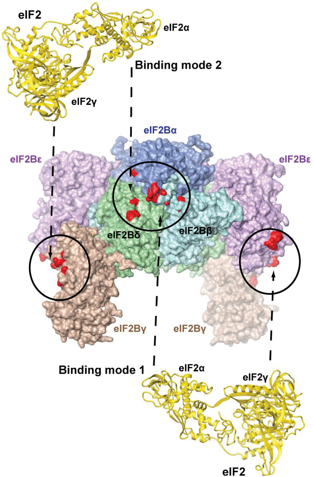
The structure of eIF2B is shown in surface representation. The individual eIF2B subunits are labeled. Sites of cross-linking to eIF2γ (left and right) and eIF2α (center) and are colored red and circled. The second eIF2α-binding pocket (not visible) is on the opposite face of the complex. The two alternative binding modes of eIF2 (shown in gold ribbon), involving the visible eIF2α-binding pocket on the front, are illustrated with dashed arrows above and below the eIF2B structure, and numbered as in the text. Note that the same two alternative eIF2 binding modes are possible on the opposite face of eIF2B, but not shown for clarity.