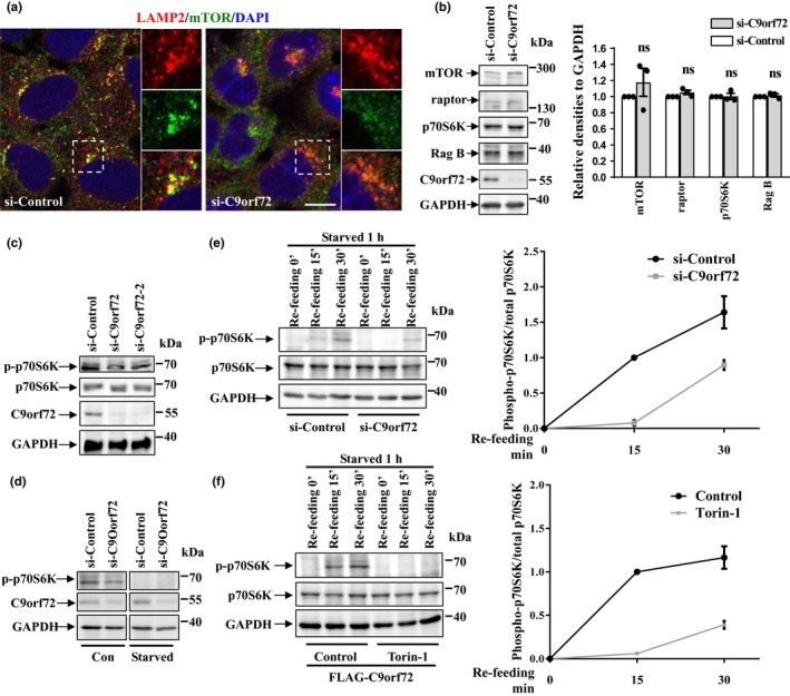
Figure 2.

Effect of C9orf72 on mTOR localization and mTORC1 activity. (a) HEK 293 cells were transfected with either control or C9orf72 siRNA for 72 hr, and then, the cells were subjected to immunofluorescence assay by antibody against LAMP2 (red) and mTOR (green). DAPI (blue) was used for nuclear staining. Scale bar, 5 µm. (b) HEK 293 cells were transfected as in A. After 72 hr, the cells were lysed and lysates were subjected to immunoblot assay using indicated antibodies. Statistical analysis from three independent experiments was presented as means ± SEM, ns, not significantly different; one‐way ANOVA. (c) HEK 293 cells were transfected with the indicated siRNAs for 72 hr. Then, the cell lysates were subjected to immunoblot analysis. (d) HEK 293 cells were transfected with the indicated siRNAs for 72 hr. Then, the cells were then incubated with Earle's balanced salt solution for 1 hr. The cell lysates were then subjected to immunoblot analysis. (e) HEK 293 cells were transfected with the indicated siRNAs for 72 hr. Then, the cells were incubated with EBSS for 1 hr and re‐fed with full medium for 15 or 30 min. The cell lysates were subjected to immunoblot analysis with indicated antibodies. The statistical analysis of relative densities is shown on the right side as means ± SD. (f) HEK 293 cells were transfected with FLAG‐tagged C9orf72. Twenty‐four hours later, the cells were incubated with EBSS for 1 hr or re‐feeding with amino acids for the indicated time, with or without Torin‐1 (250 nM) treatment. The statistical analysis of relative densities is shown on the right side as means ± SD
