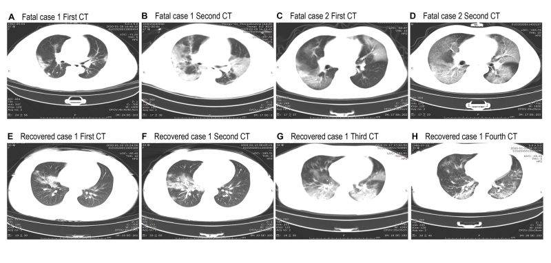
Fig 1.
Representative chest computed tomographic images of patients with covid-19 who died and patients who recovered. A-D are chest computed tomograms showing axial view lung window from two deceased patients. Case 1 was a 57 year old women, and case 2 was a 53 year old man. E-H are chest computed tomograms images from a 33 year old woman who recovered. A: image obtained on day 10 after symptom onset shows multiple ground glass opacities and consolidation in bilateral lungs. B: image obtained on day 18 after symptom onset shows progressive multiple ground glass opacities and consolidation in bilateral lungs. C: image obtained on day 9 after symptom onset shows multiple ground glass opacities in bilateral lungs and solid nodule in right lower lobe. D: image obtained on day 13 after symptom onset shows progressive ground glass opacities in bilateral lungs and decreased density of solid nodule in right lower lobe. E: image obtained on day 4 after symptom onset shows right middle lobe and lower lobe consolidation and ground glass opacities. F: image obtained on day 7 after symptom onset shows progressive right middle lobe and lower lobe consolidation and ground glass opacities. G: image obtained on day 11 after symptom onset shows progressive multiple ground glass opacities and consolidation in bilateral lungs and decreased density and range of right middle lobe consolidation. H: after 17 days’ therapy, follow-up computed tomograms show ground glass opacities, and consolidation are obviously resolved in bilateral lungs

