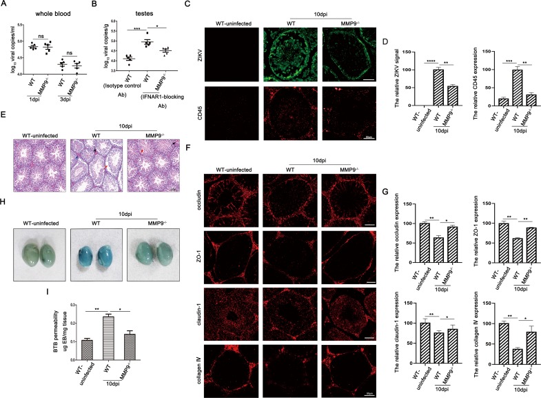
Fig 2. MMP9 compromised BTB integrity to facilitate ZIKV entry into the testes.
C57BL/6 WT and MMP9-/- male mice (6–7 weeks old) treated with Ifnar-blocking mouse monoclonal antibodies were infected intraperitoneally with ZIKV (1 × 10⁷ PFU). The C57BL/6 WT mice treated with isotype control antibodies were also infected intraperitoneally with ZIKV (1 × 10⁷ PFU) as a mock control. (A) RNA was extracted from the whole blood, and a probe-based assay was used to quantify viral RNA copy number by TaqMan qPCR amplification of ZIKV E gene at different time points. Data shown are means ± SEMs; ns, not significant, n = 5. (two-tailed Student’s t-tests). (B) RNA was extracted from the testes, and a probe-based assay was used to quantify viral RNA copy number by TaqMan qPCR amplification of ZIKV E gene at different time points. Data shown are means ± SEMs; *P< 0.05; **P< 0.01; ***P< 0.001, n = 5. (one-way ANOVA). (C and D) Results of immunofluorescence staining for ZIKV (green) and CD45 (red) in the testes and the quantifications were shown using Image J. Data shown are means ± SEMs; *P< 0.05; **P< 0.01; ***P< 0.001, ****P< 0.0001, n = 5 (one-way ANOVA), Scale bar, 50μm. (E) Histopathological changes in the testes of WT and MMP9-/- mice on day 10 post infection. Disrupted seminiferous tubules with leukocyte infiltration (blue arrow), abnormally organized cells (red arrow), and intratesticular congestion (black arrow) were obviously observed in ZIKV-infected WT testes. Representative images from several independent experiments are shown, Scale bar, 100μm. (F and G) Results of immunofluorescence staining of occludin (red), ZO-1 (red), claudin-1 (red), and type Ⅳ collagen (red) and the quantifications for the percentage of these proteins were shown using Image J. Data shown are means ± SEMs; *P< 0.05; **P< 0.01, n = 5 (one-way ANOVA), Scale bar, 50μm. (H) Evans blue BTB permeability. On day 10 postinfection, WT and MMP9-/- mice were injected with Evans blue and perfused 1h later. Uninfected WT mice were used as a control. Data are representatives of the results of three experiments (n = 8/group). (I) Quantification of Evans blue in the mouse testes. Evans blue was extracted from whole testes, and absorbance was measured, using uninfected-mouse testis extracts as a blank. *P< 0.05; **P< 0.01. ns, not significant (one-way ANOVA).

