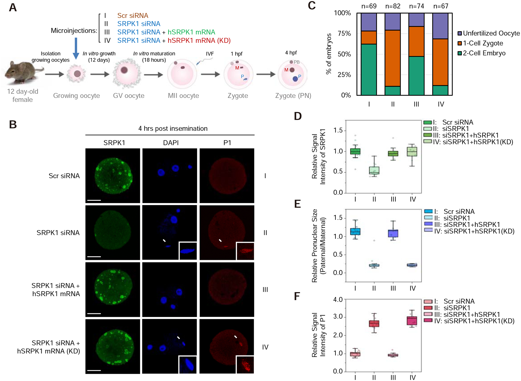
Figure 2. Kinase activity of SRPK1 is required for protamine removal.

(A) Scheme for siRNA microinjection into growing oocytes to deplete maternal SRPK1 and functional rescue with siRNA-resistant SRPK1 mRNA. See Figure S2A for characterization of the growing oocytes.
(B) Representative images of fertilized eggs stained with anti-SRPK1 (green), anti-P1 (red), and DAPI (blue) 4 hours post insemination. Panel I to IV respectively show oocytes injected with scrambled (Scr) siRNA (I), SRPK1 siRNA (II), SRPK1 siRNA plus human SRPK1 mRNA (III) and SRPK1 siRNA plus human kinase-dead SRPK1 mRNA (KD) (IV). Arrows indicate paternal DNA with a zoomed image in the insert. P, paternal DNA; M, maternal DNA; PB, polar body. Scale bar, 20 μm.
(C) Quantified impact on zygotic development in response to different treatments as in B. The numbers of zygotes quantified from three independent experiments are indicated above the bars. See Figure S2B for additional details.
(D,E,F) Relative SRPK1 staining signals (D), relative values of pronuclear size (paternal/maternal) (E), and relative P1 staining signals (F) in fertilized eggs. The values of zygotes treated with Scr siRNA were set as 1.0 in each case. **P < 0.01 by two-tailed Student’s t-test; error bars, mean±SEM.
