
Figure 6. Phosphorylation-dependent interactions of Protamine 1 with NPM and HIRA.
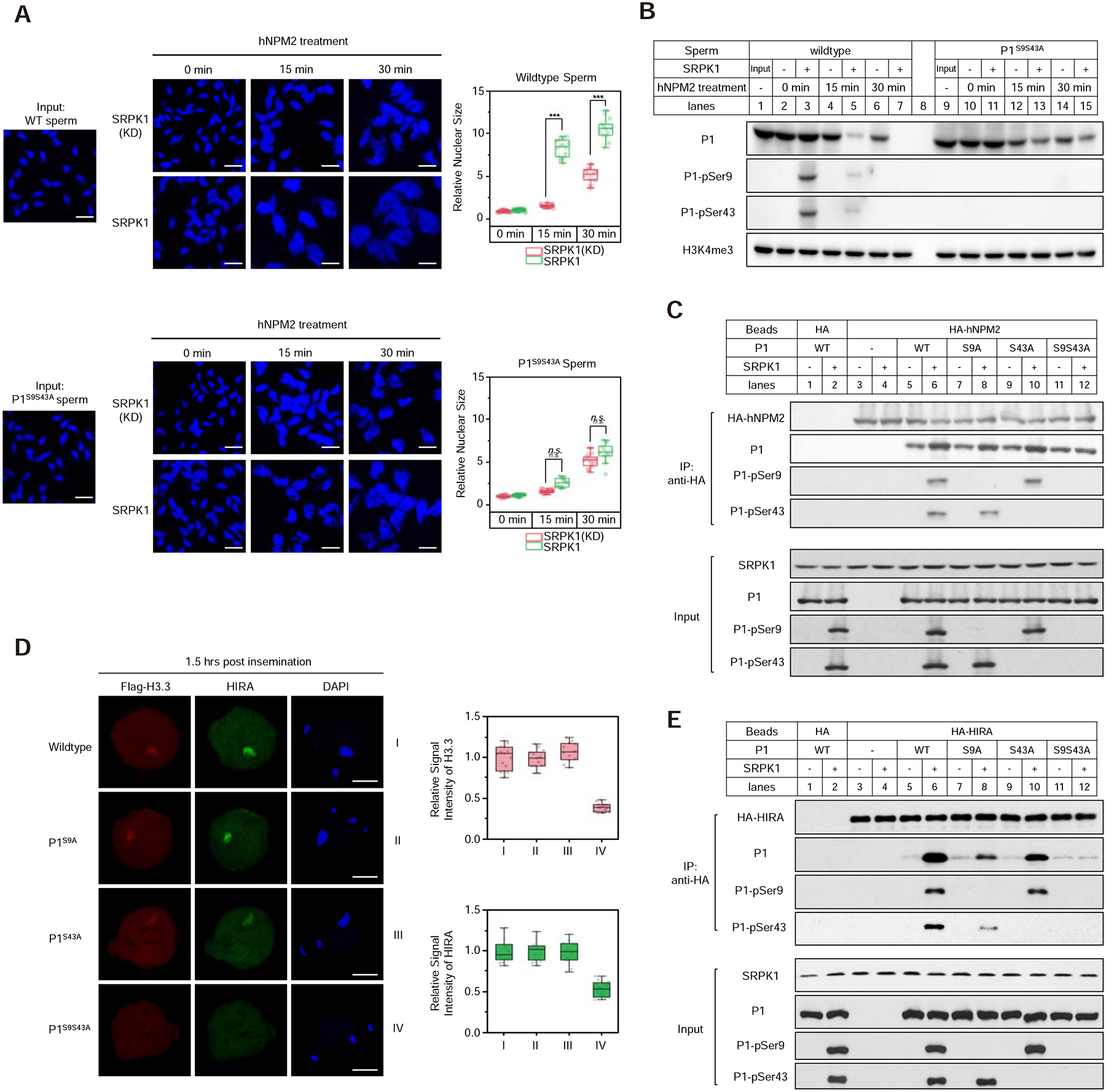
(A) SRPK1 phosphorylation-dependent enhancement of sperm DNA decondensation with purified NPM2. Isolated wild-type (upper panels) and double mutant (lower panels) sperm were treated with purified NPM2 for different time points in the presence of kinase-dead or active SRPK1. Sperm volumes were quantified, as shown on the right. ***P < 0.001 by two-tailed Student’s t-test; error bars, mean±SEM. See Figure S6A for the in vitro sperm DNA decondensation assay.
(B) Western blot of retained P1 on sperm chromatin from the experiments as in A. Total and specifically phosphorylated P1 were blotted with specific antibodies. See Figure S6B for purification of recombinant NPM2, Figure S6C and S6D for the levels of retained P1 upon longer NPM2 or SRPK1 treatment, Figure S6E and S6F for in vivo results on paternal DNA decondensation in SRPK1-depleted and P1 double mutant oocytes ~10-hour post insemination, and Figure S6G for the lack of effect of NPM2 on in vitro assembled P1/DNA particles.
(C) Pulldown assay to determine the interaction between HA-tagged human NPM2 and P1 in the presence or absence of SRPK1. Anti-HA pulldowns were blotted with individual antibodies as indicated.
(D) Representative images of fertilized eggs stained for FLAG-H3.3 (red), HIRA (green) and DNA (blue) 1.5-hour post insemination with wild-type, single and double mutant sperm. P, paternal DNA; M, maternal DNA; PB, polar body. Scale bar, 20 μm. Fluorescence intensity was quantified by ImageJ, as shown on the right for FLAG-H3.3 and HIRA.
(E) Pulldown assay to determine the interaction between HA-tagged human HIRA and P1 in the presence or absence of SRPK1. Anti-HA pulldowns were blotted with individual antibodies as indicated.