
Fig. 3. Phenotypic evaluation of macrophages (MΦs) carrying IFN-γ backpacks in vitro.
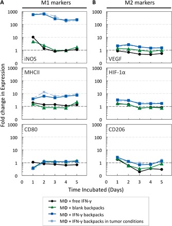
BMDMs were cultured for 5 days with free IFN-γ (16 ng/ml; black lines), blank backpacks (0 ng/ml IFN-γ; green lines), and IFN-γ backpacks (16 ng/ml equivalent) in normoxia (dark blue lines) and tumor-mimicking conditions (1% O2 and 10 volume % tumor-conditioned media; light blue lines). Cellular expression of representative (A) M1 markers (iNOS, MHCII, and CD80) and (B) M2 markers [vascular endothelial growth factor (VEGF), hypoxia-inducible factor 1α (HIF-1α), and CD206], relative to that of unpolarized macrophages (without IFN-γ or backpacks). Graphs are logarithmic (n = 10,000 events per data point).