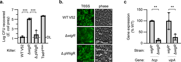
Figure 5: WigR regulates the T6SS in V52.
a. Relative survival of E. coli prey after killing by V52 with intact (WT), deleted (Δ), or plasmid complemented (Δ,p) wigR. One-way ANOVA with Tukey’s test; ***, p < 0.001. DL, detection limit. b. Phase contrast (right) and VipA-GFP T6SS assembly (left) in V52 with intact (WT), deleted (Δ), or complemented (Δ,p) wigR. Representative of three independent replicates. Scale bar, 2 μm. c. Expression of main (vipA) and auxiliary (hcp) T6SS operons in wild-type (wigR+) or ΔwigR V52. One-way ANOVA with Sidak’s test; **, p < 0.01. Graphs show the mean of at least three independent replicates, error bars show standard deviation. Dots show individual replicates.

