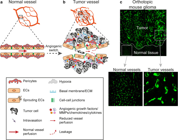
Fig. 2.
Morphological and functional characteristics of tumor vessels as compared to normal vessels. a Normal vessels display an organized and hierarchical branching pattern of arteries, veins, and capillaries. In healthy vessels, endothelial cells are supported by basal membrane and pericytes coverage and they are tightly connected by stable cell-cell junctions. b Tumor vessels are morphologically and functionally different from normal vessels. In response to persistent and imbalanced expression of angiogenic factors and inhibitors, tumor vessels display an unorganized network lacking of a hierarchical vessel division. Tumor vessels are characterized by reduced blood flow, endothelial cell sprouting, disruption of endothelial cell junctions, loss of pericytes coverage and increased vessel leakiness resulting in increased tissue hypoxia and intravasation of tumor cells. Moreover, tumor endothelial cell basal membrane is abnormal, including loose associations with endothelial cells and variable thickness. c Tumor vessel abnormalization shown by immunofluorescent staining for the vessel marker CD31 (green) in an orthotopic syngeneic mouse model of glioma growing in the brain

