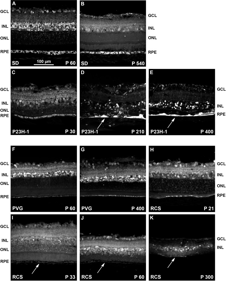
Figure 8.
Intraretinal fluorogold tracing shows the retinal remodeling of the dystrophic strains. (A–E) Magnifications from retinal cross-sections showing fluorogold tracing in young (A) and old (B) SD control rats, and 30 (C), 210 (D) and 400 (E) days old P23H-1 rats. (F–K) Magnifications from retinal cross-sections showing fluorogold tracing in young (F) and old (G) PVG control rats, and 21 (H), 33 (I), 60 (J) and 300 (K) days old RCS rats. In the P23H-1 retinas, aberrations in the retinal layered structure, and the RPE morphology (white arrows in D,E) are observed from P210. The RCS retina degenerates earlier, and at P60 the ONL and the RPE have almost disappeared with only some isolated RPE cells remaining (white arrows in I-K). P: postnatal day. GCL: ganglion cell layer. INL: inner nuclear layer. ONL: outer nuclear layer. RPE: retinal pigment epithelium.

