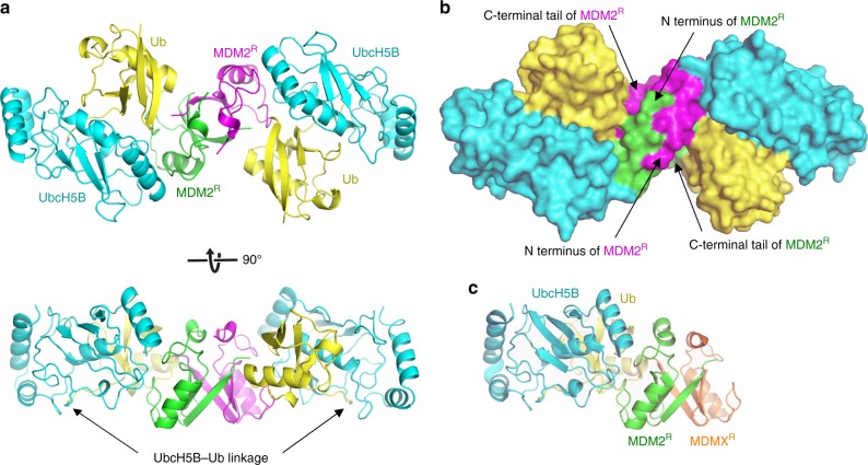
Fig. 2. Crystal structure of human MDM2 RING domain homodimer bound to UbcH5B–Ub.
a Cartoon representation of the complex. One MDM2R is colored green and the other is in magenta. UbcH5B is in cyan and Ub is in yellow. UbcH5B–Ub linkage is indicated. Top and bottom panels are related by 90° rotation about the x-axis. b Surface representation of the complex, colored and oriented as in a (top panel). The N-terminal region preceding the RING domain and the C-terminal tail of MDM2 are indicated. c A cartoon representation of the structure of MDM2-MDMX RING domain heterodimer bound to UbcH5B–Ub (PDB ID: 5MNJ) oriented in the same view as in a (bottom panel). MDM2R, UbcH5B, and Ub are colored as in a and MDMX RING domain (MDMXR) is in orange.

