Figure 2.
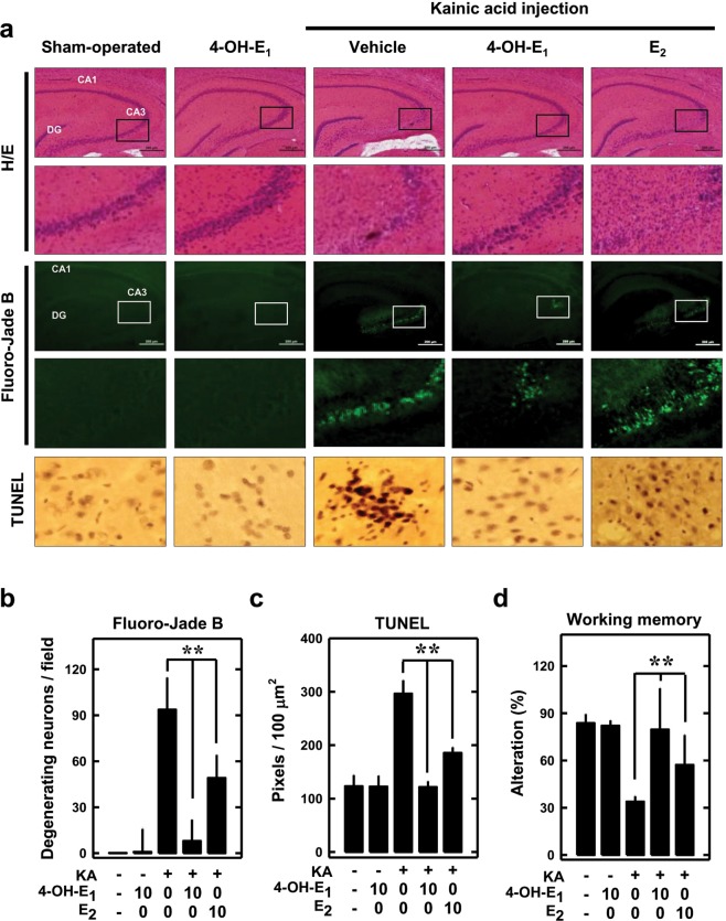
4-OH-E1 attenuates kainic acid (KA)-induced neurodegeneration in rat hippocampus in vivo. (a) KA (1 µL of 1 µg/µL solution) is injected into the left and right lateral ventricles of male rats (anterior/posterior, ‒1.0; rostral, ±1.6; dorsal/ventral, 4.5) using a microliter syringe under anesthesia with ketamine and xylazine (50 and 5 mg/kg, s.c.). The control rats (sham-operated) are injected with 1 µL of saline. Immediately following kainic acid injection, the animals receive daily s.c. administration of 10 µg/rat of 4-OH-E1 or E2 for 7 consecutive days. Control rats receive the same s.c. administrations of vehicle only. Shown are representative data for the histopathological analysis (H/E staining) of the hippocampal region (H/E, upper panels, 40×) and the enlarged CA3 region (H/E, lower panels); the Fluoro-Jade B staining of the hippocampal region (Fluoro-Jade B, upper panels) and the enlarged CA3 region (Fluro-Jade B, lower panels); and the TUNEL staining of apoptotic cells (TUNEL). (b,c) Quantitative data for the number of degenerating neurons (b) and TUNEL-positive cells (c) in control and kainic acid-treated rats. (d) Working memory in control and kanic acid-treated rats based on the Y-maze test (conducted 6 days after kainic acid treatment). For all quantitative data, each value is the mean ± SD (n = 5). *P < 0.05 versus the KA alone group.

