
Fig. 1.
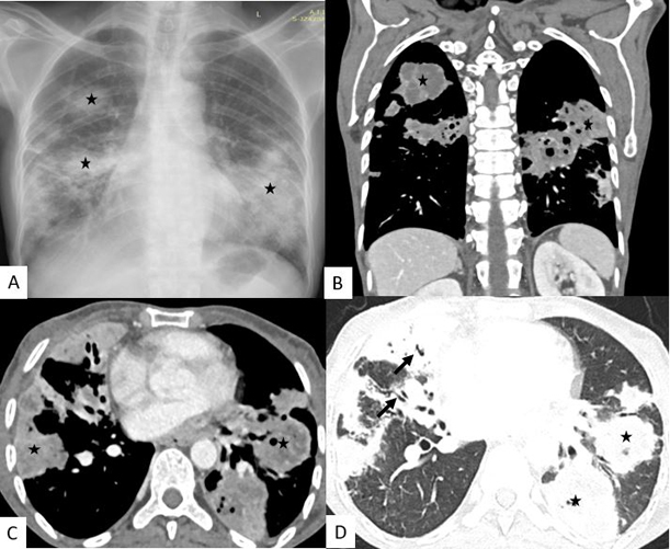
Chest Radiograph posteroanterior view (A) showing multifocal patchy consolidation (asterix) in bilateral lower, mid and right upper zone. Contrast Enhanced Computed tomography (CECT) of thorax coronal (B) and axial (C) with showing multiple consolidation (asterix) in bilateral lower and right middle and upper lobes with internal hypodensities suggestive of necrosis. Lung window axial sections (D) also showing similar multiple consolidations with internal air-bronchogram (black arrow).