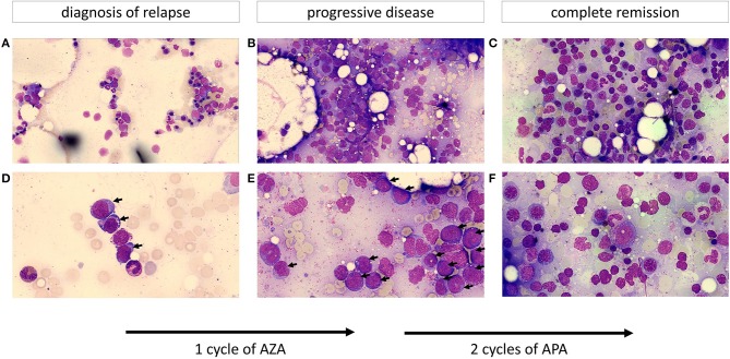
Figure 1.
Cytomorphology of bone marrow aspirates (A–C depict an overview of the slide; D–F show the cells at greater magnification; objective 63x). (A,D) Show a hypoplastic bone marrow after stem cell transplantation with up to 10% myeloblasts (marked by an arrow) resembling early relapse after allo-HSCT. Leukemic blasts display a basophilic cytoplasm frequently containing vacuoles. (B,E) (after one cycle of AZA) demonstrate a hypercellular marrow with ~50% of the previously described leukemic blasts (progressive disease). (C,F) (after two cycles of APA) show a normocellular marrow with an increased and left-shifted erythropoiesis, differentiated neutrophils and no evidence of an increased percentage of myeloblasts resembling complete remission.

